Definition of "Angiography"
Last modified: 15 hours
It's where you make an image of the inside of a blood vessel.
X-ray doesn't show soft tissue very well. So we need to pump contrast into the blood system, to help highlight the blood vessels.
Angiography (from Greek "angio" meaning "vessel", and "graphy" meaning "to write") is medical imaging to visualize the lumen (inside) of blood vessels and organs of the body, with particular interest in the arteries, veins and heart chambers. Angiogram (aka angiograph) is the film/image of the blood vessel. Angiogram is usually used synonymously with arteriogram, and the word venogram used more precisely.
Patient information
What is angiography?It's where you make an image of the inside of a blood vessel.
Method
- Injecting a radio-opaque contrast agent into the blood vessel and imaging using x-ray based techniques (e.g. fluoroscopy)
- Depending on the type of angiogram, access to the blood vessels is gained most commonly through the:
- Femoral artery, to look at the L side of the heart and at the arterial system
- Jugular or femoral vein, to look at the R side of the heart and the venous system
- Using a system of guide wires and catheters, a type of contrast agent (which shows up by absorbing the x-rays), is added to the blood to make it visible on the x-ray images
- X-ray images taken may either be still images displayed on an image intensifier or film, or as a movie (motion images)
- Digital subtraction angiography (DSA) is the technique used to take for all structures except the heart, which involves taking 2-3 frames per second, allowing the radiologist to evaluate the flow of the blood through vessel(s). This technique "subtracts" the bones and other organs so only the vessels filled with contrast agent can be seen
- Because DSA requires the Pt to remain motionless, it can't be used for the heart. Heart images are taken at 15-30 frames per second, not using a subtraction technique
- The techniques can allow a cardiologist to see stenosis (blockages/narrowings) inside the vessel, which may inhibit the flow of blood, and cause pain
Patient information
How do you take pictures of the inside of a blood vessel, when it's inside your body? Do you use an x-ray?X-ray doesn't show soft tissue very well. So we need to pump contrast into the blood system, to help highlight the blood vessels.
Classification
- Coronary angiography, one of the most common angiograms, performed to visualize the blood in the coronary arteries. A catheter (long, thin, flexible tube) is used to administer the x-ray contrast agent at the desired area to be visualized. The catheter is threateded into an artery in the forearm, and the tip is advanced through the arterial system into the major coronary artery. X-ray images of the transient radiocontrast distribution within the blood flowing inside the coronary arteries allowing visualization of the size of the artery openings. Presence/absence of atherosclerosis or atheroma within the walls of the arteries can NOT be clearly determined. CT is better than MRI to detect coronary artery disease, with both sensitivity/sepcificty, cheaper, and shorter breath-hold time
- Microaniography, used to visualize tiny blood vessels
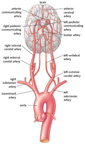
- Neurovascular [digital subtraction] angiography, used to visualize the arterial and venous supply to the brain. Intervention work e.g. coil-embolization of aneurysms and AVM gluing can also be performed. This includes imaging of the Circle of Willis (aka cerebral arterial circle), which can be imaged together with the arch of aorta
Source: Class Connection
- Peripheral angiography, done routinely through the femoral artery, but can also be performed through the brachial or axillary/arm artery. Any stenosis found may also be Tx using atherectomy. Peripheral angiography is performed to identify:
- Vessel stenosis (narrowing) in Pt's w/ leg claudication or cramps, caused by reduced blood flow down the legs and to the feet
- Pt's w/ renal stenosis, which commonly causes HTN
- Used in the head to find and repair stroke
- Post-mortem CTA for medicolegal cases
- Cholangiography, which is imaging of the bile duct (aka biliary tree) by x-rays. In both cases, fluorescent fluids are used to create contrasts that make the Dx possible. It has replaced the previously used method of intravenous cholangiography. It includes:
- Percutaneous transhepatic cholangiography (PTC), examination of liver and bile ducts by x-rays. This is done by insertion of a thin needle into the liver carrying a contrast medium to help see a blockage in the liver and bile ducts
- Endoscopic retrograde cholangiopancreatography (ERCP), although this is a form of imaging, it is both Dx and Tx, and often classified with surgeries rather than imaging
- Although the term is strictly defined as based on projectional radiography (i.e. based on x-rays), it has been applied to newer vascular imaging techniques (e.g. CT angiography and MR angiography)
- Isotope angiography, more correctly refers to an isotope perfusion scan
Complications
- Risk of heart attack is actually narrowed down, as heart strength doubles after an angiogram surgery. A sudden shock can cause little pain at the surgery area, but heart attacks and strokes usually don't occur, like in bypass surgery
Complications of cerebral angiography (e.g. digital subtraction angiography, or contrast MRI) include:
- Bleeding or bruising at the site where the contrast is injected
- Stroke
- Allergic reaction to the anesthetic or contrast medium
- Blockage or damage to one of the access veins in the leg
- Thrombosis and embolism formation
- Delayed bleeding
See also
- X-ray
- CT
Synonyms:
Angio
Angiogram
Angiograph
Angiographic
Arteriogram
Arteriograph
Cerebral arterial circle
Cholangiogram
Cholangiograph
Cholangiography
Circle of Willis
Coronary angiography
Isotope angiography
MIcroaniography
Neurovascular angiography
Neurovascular digital subtraction angiography
Percutaneous transhepatic cholangiography
Peripheral angiography
PTC
Venogram
Venograph
Find a practitioner
Practitioner count: 0
Sponsor a disease. And see how your proceeds help.
$1
Express interest
$10
Write text
$40
Write FAQ
$100
Snap photos
$400
Record audio
$1k
Produce video
$4k
Interview experts